
The Ultimate Guide to Swedish Bodywork Benefits
October 1, 2026
The Ultimate Guide to the Best Hair Massage Oil for Your Scalp
October 6, 2026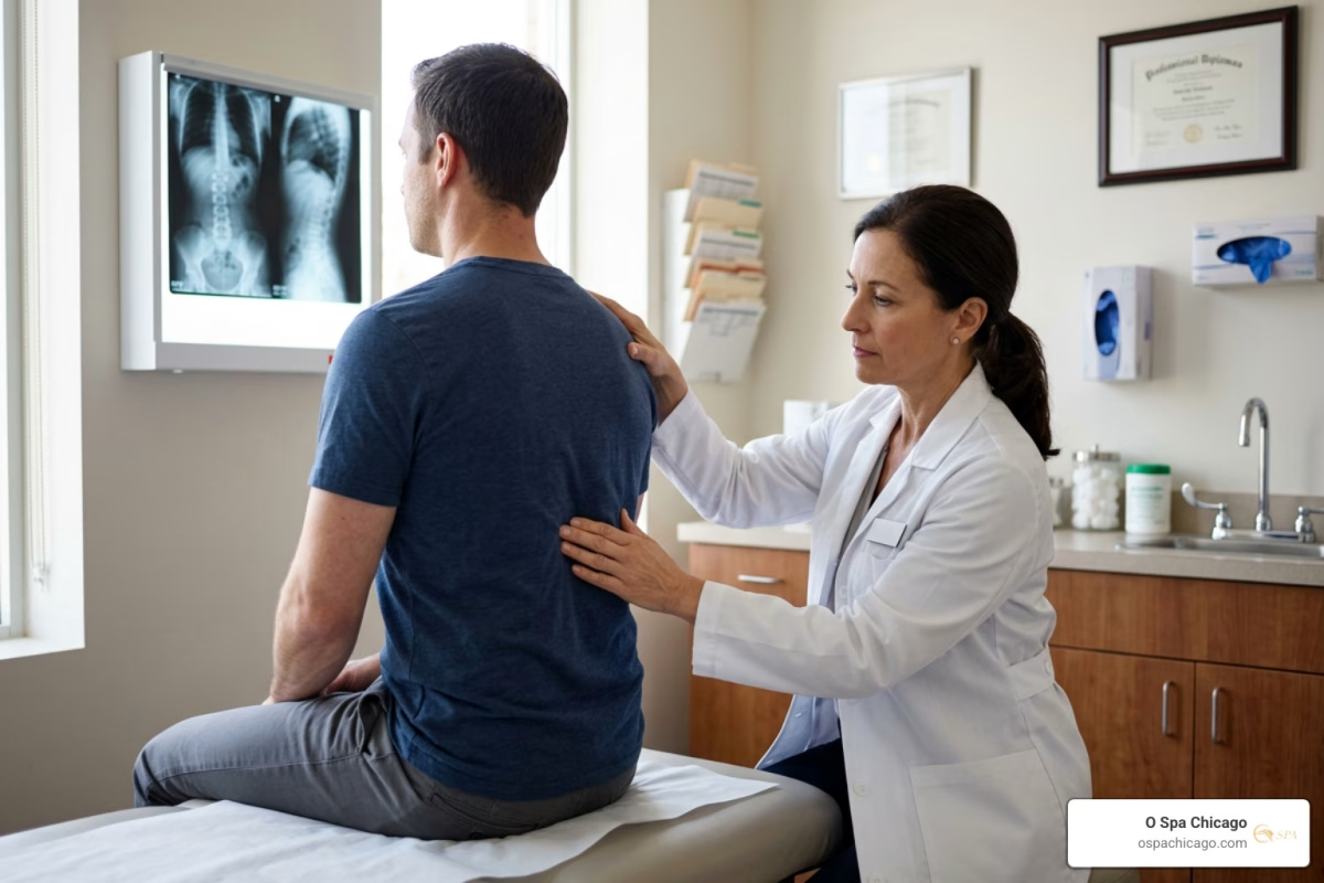
Fractured Back Treatment: Start With Safety, Then Choose the Right Care
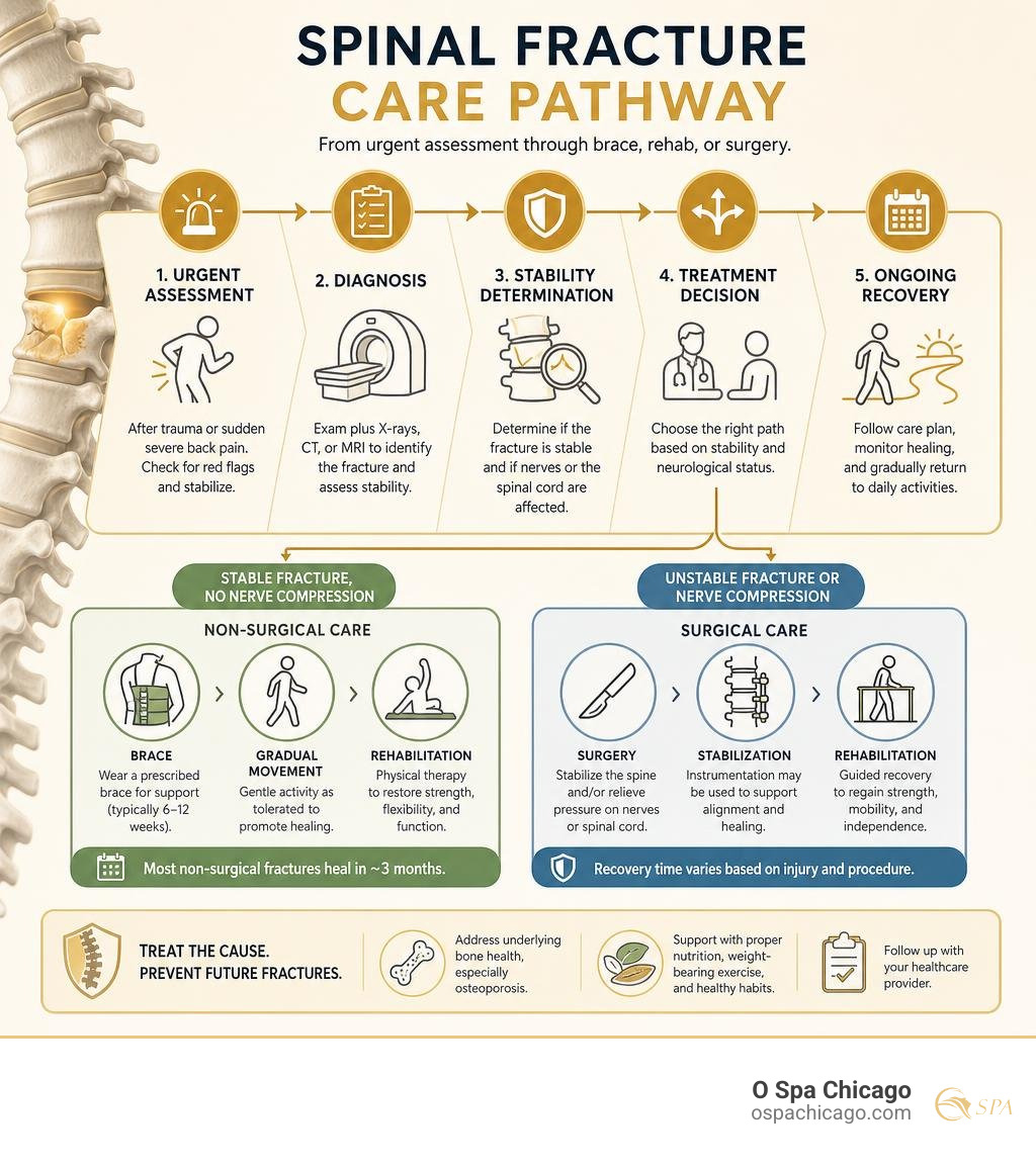
Fractured back treatment depends on whether the broken vertebra is stable and whether nerves or the spinal cord are affected. After a fall, crash, or sudden severe back pain, get medical care promptly. Doctors typically use an exam plus X-rays, CT, or MRI to identify the fracture. Stable fractures often heal with pain control, a brace, gradual movement, and physical therapy. Unstable fractures or fractures causing nerve pressure may need surgery.
Seek emergency help now for back pain after major trauma, new weakness or numbness, trouble walking, or loss of bladder or bowel control. You may be able to walk with some spinal fractures, but that does not prove the injury is minor.
A “broken back” can mean anything from an osteoporotic compression fracture to a severe burst fracture. Most fractures that do not need surgery heal in roughly three months, while many compression-fracture patients wear a prescribed brace for six to twelve weeks. The right plan also treats the cause, especially osteoporosis, because one compression fracture greatly raises the risk of another.
I am June Liu, a Chicago wellness professional and owner of O Spa Chicago, with experience helping clients understand supportive self-care, stress relief, and recovery-minded wellness. While fractured back treatment requires medical diagnosis and direction from a qualified spine clinician, this guide explains the care path in clear, practical terms.
Understanding Spinal Fractures: Types, Causes, and Diagnosis
The adult human spine consists of 33 interlocking vertebrae divided into distinct anatomical regions: the cervical spine (neck), thoracic spine (mid-back), lumbar spine (lower back), sacrum, and coccyx. These bony segments shield the delicate spinal cord while providing structural support for upright posture and flexible movement. According to clinical overviews of vertebral fractures, a spinal fracture occurs whenever excessive axial loading, sudden rotation, or underlying bone disease causes a structural crack or collapse in one or more vertebrae.
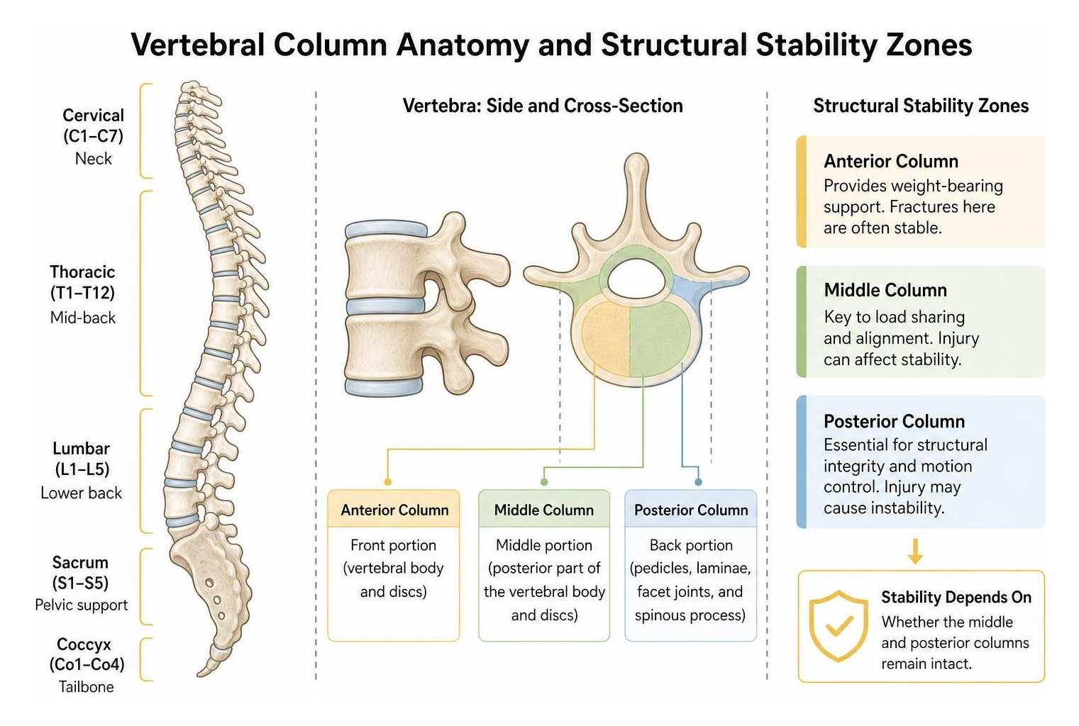
Spinal fractures vary significantly depending on whether the middle and posterior columns of the vertebra remain intact. When a break compromises the structural alignment of the spinal canal, fragments can press against the spinal cord or exiting nerve roots, transforming an isolated orthopedic injury into a complex neurological condition.
Common Causes and Osteoporosis Risks
Vertebral fractures generally stem from two distinct mechanisms: high-energy trauma or low-energy osteoporotic fragility. In the United States alone, high-impact incidents such as motor vehicle collisions, falls from significant heights, and severe sports collisions cause more than 150,000 spinal fractures annually.
Conversely, bone-density loss represents the leading driver of non-traumatic vertebral collapse. Osteoporosis weakens the internal trabecular microarchitecture of bone, leaving vertebrae vulnerable to fractures during everyday activities such as bending over, lifting groceries, or coughing.
- Over 1.5 million vertebral compression fractures occur each year in the U.S. due to osteoporosis.
- Approximately 1 in 4 females with an osteoporotic compression fracture never receive a formal diagnosis.
- Sustaining a single osteoporotic compression fracture increases the statistical risk of developing a subsequent vertebral fracture by five times.
Fracture Classifications: Stable vs Unstable Patterns
Spine specialists classify vertebral injuries based on the direction of applied force, the specific column of the spine damaged, and overall mechanical stability, as detailed in the AAOS thoracic and lumbar spine fracture guide.
- Compression Fractures: Typically caused by flexion forces, the anterior (front) wall of the vertebral body collapses under pressure while the middle and posterior bony walls remain intact. These are generally stable and rarely compromise the spinal cord.
- Burst Fractures: A high-energy vertical force crushes the vertebral body entirely, causing bone fragments to spread outward in multiple directions. If fragments retropulse into the spinal canal, they can compress nerve structures.
- Chance Fractures (Flexion-Distraction): Often seen in motor vehicle collisions where a lap-only seatbelt holds the pelvis while the torso thrusts violently forward, pulling the vertebra and its posterior ligaments apart.
- Fracture-Dislocations: Severe, high-energy rotational or shearing injuries that disrupt both bone and supporting ligaments, frequently resulting in unstable displacements and neurological deficits.
Diagnostic Protocols and Imaging Tests
An accurate diagnosis begins with a comprehensive physical and neurological exam to assess motor strength, sensation, and deep tendon reflexes. Spine specialists then employ targeted imaging to categorize the break:
- Plain Radiography (X-rays): Serves as the primary frontline screening tool to evaluate vertebral alignment, detect overt deformities, and calculate height loss.
- Computed Tomography (CT) Scans: Considered the gold standard for bone detail, CT scans deliver cross-sectional views that confirm whether fracture fragments extend into the spinal canal.
- Magnetic Resonance Imaging (MRI): Essential for assessing soft-tissue integrity, spinal cord edema, intervertebral disc disruption, and distinguishing acute fractures from older, healed bone collapse.
- Dual-Energy X-ray Absorptiometry (DEXA): Measures bone mineral density to evaluate underlying osteopenia or osteoporosis, establishing whether long-term metabolic therapy is needed, consistent with an evidence-based vertebral fracture evaluation (Whitney & Alastra, 2023).
Non-Surgical Fractured Back Treatment: Bracing and Conservative Care
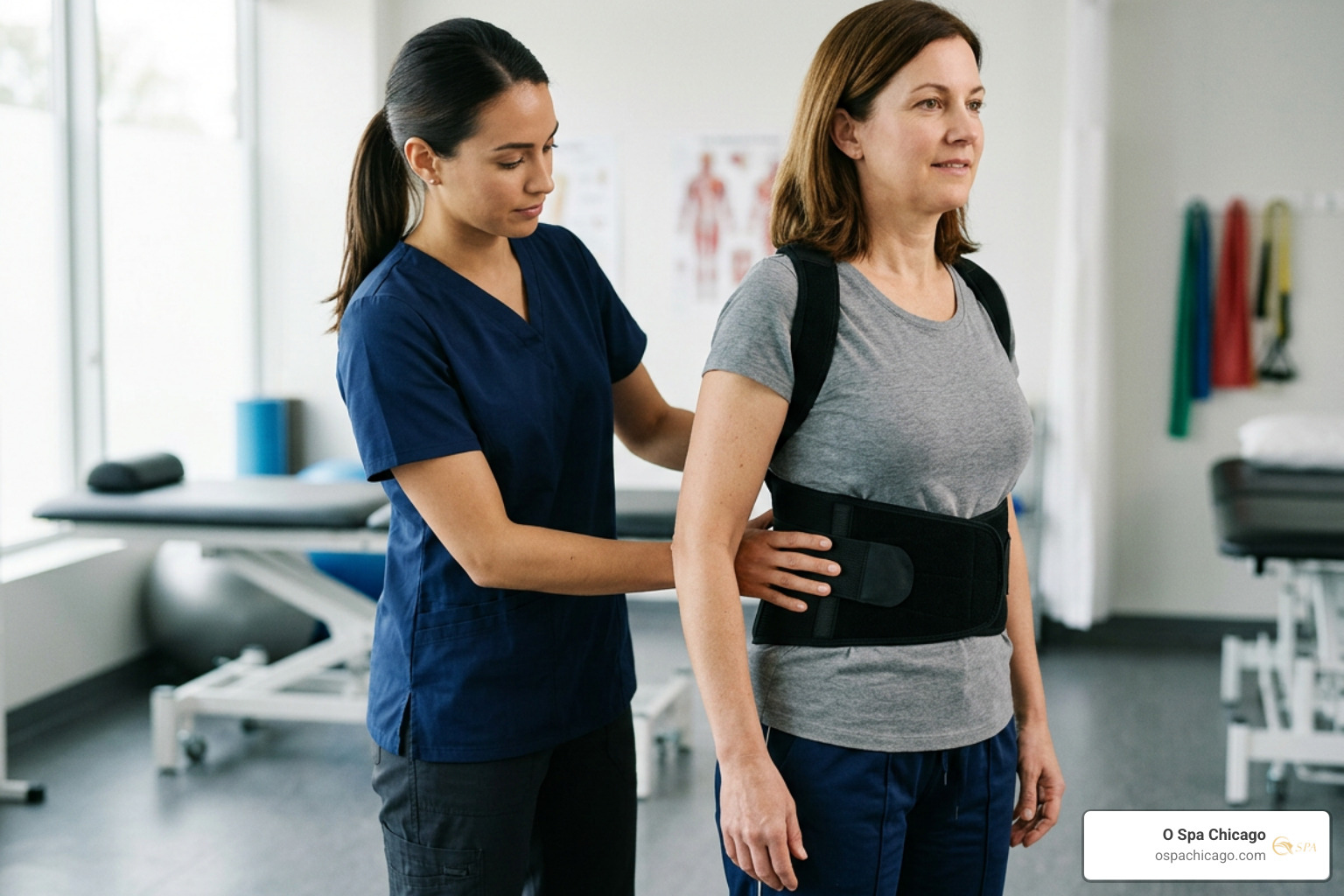
When diagnostic imaging confirms a stable spinal fracture without neurological compromise, non-surgical conservative care serves as the preferred frontline medical intervention (Genev et al., 2017). The goal of non-operative management is to control pain, maintain spinal alignment, and support natural bone healing.
| Treatment Approach | Typical Indication | Primary Mechanism | General Recovery Window |
|---|---|---|---|
| Conservative Care | Stable compression fractures, minor burst fractures | External bracing, pain medications, physical therapy | 6 to 12 weeks of bracing; ~3 months for bone consolidation |
| Kyphoplasty / Vertebroplasty | Painful osteoporotic VCFs refractory to rest | Minimally invasive cement injection with/without balloon cavity | Immediate pain relief; light routine resumption in 1 to 2 weeks |
| Spinal Fusion Surgery | Unstable fractures, severe burst injuries, neurological compression | Pedicle screw fixation, rods, and bone grafting with decompression | 6 weeks initial surgical healing; 6 to 12 months full fusion remodeling |
Bracing Protocols and Healing Timelines for Fractured Back Treatment
External orthotic braces act as a supportive cast for the torso, limiting flexion, extension, and lateral rotation to allow fractured vertebral trabeculae to bridge and remodel. Depending on the level of the injury, clinicians prescribe specific orthoses:
- Thoracolumbosacral Orthosis (TLSO): A custom-molded rigid plastic shell that stabilizes fractures occurring between the mid-thoracic spine and the upper lumbar segments.
- Hyperextension Braces (e.g., Jewett or CASH): Lightweight metal-framed braces designed to prevent forward bending, keeping the spine in slight extension to offload pressure from the anterior vertebral body.
Most patients wear a prescribed brace for six to twelve weeks during upright activities. Non-operative fractures generally achieve solid radiographic healing within three months, during which patients are advised to avoid heavy lifting, strenuous twisting, and prolonged sitting without spinal support.
Medication and Pain Management Strategies
Pharmacologic treatment aims to manage acute discomfort so patients can mobilize safely without prolonged bed rest:
- Short-Term Analgesics and NSAIDs: Over-the-counter or short courses of prescription anti-inflammatory medications help mitigate acute muscular and osseous pain.
- Calcitonin Salmon: A subcutaneous or nasal-spray peptide that can reduce acute osteoporotic bone pain while inhibiting osteoclast activity.
- Anti-Resorptive and Anabolic Agents: Medications such as bisphosphonates (alendronate, zoledronic acid) or teriparatide help rebuild bone microarchitecture and lower the risk of subsequent fractures.
- Neuropathic Modulators: For radicular discomfort resulting from local nerve irritation, medications like gabapentin may be utilized under physician supervision.
Physical Rehabilitation and Supportive Recovery
Prolonged bed rest leads to muscular deconditioning, bone demineralization, and secondary stiffness. Once acute healing begins, guided physical therapy introduces gentle isometric core exercises, pelvic tilts, and posture training to restore spinal stability.
As bone consolidation solidifies, patients often seek complementary wellness therapies to alleviate secondary muscle tension and postural fatigue. Exploring gentle body treatments and light massage services after medical clearance can help soothe tight paraspinal muscles, enhance circulation, and support physical comfort alongside clinical rehabilitation.
Surgical Options: Vertebroplasty, Kyphoplasty, and Spinal Stabilization
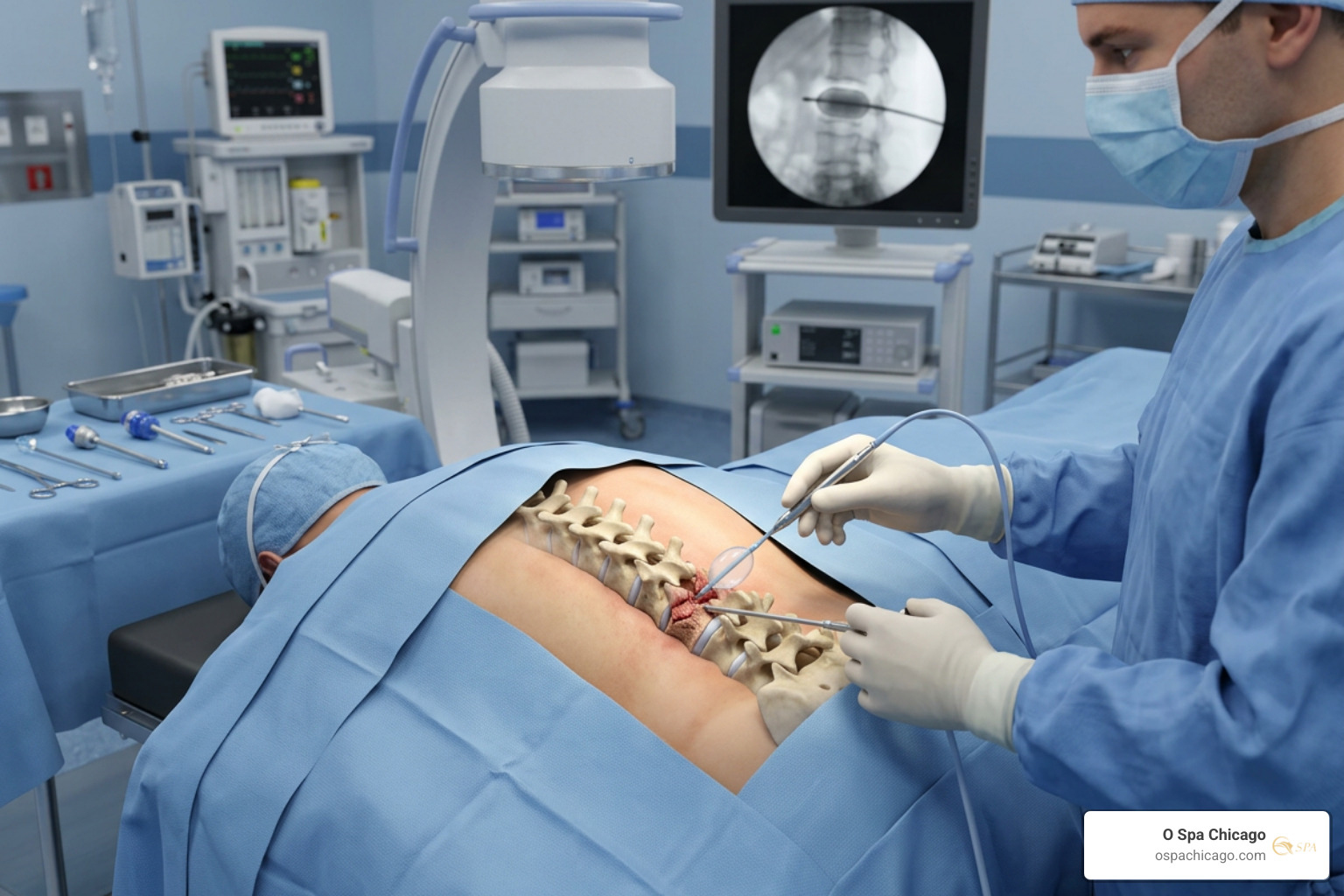
When conservative care fails to relieve debilitating pain, or when an injury disrupts the mechanical integrity of the spine, surgical intervention becomes necessary. The thoracolumbar injury classification guidelines assist orthopedic and neurosurgical specialists in balancing conservative management against surgical stabilization based on spinal canal narrowing, kyphotic deformity, and neurological status.
Vertebral Augmentation: Kyphoplasty vs Vertebroplasty
Vertebral augmentation procedures are minimally invasive outpatient treatments designed primarily for painful vertebral compression fractures (VCFs) caused by osteoporosis or osteolytic tumors:
- Percutaneous Vertebroplasty: A spine specialist inserts a hollow needle (trocar) directly into the fractured vertebral body under real-time fluoroscopic (X-ray) guidance. Low-viscosity polymethylmethacrylate (PMMA) bone cement is injected to stabilize internal microfractures and reduce painful micromotion.
- Balloon Kyphoplasty: Before cement injection, an inflatable orthopedic balloon is inserted into the compressed vertebra and inflated. This creates a controlled cavity and can partially restore lost vertebral height before the cavity is filled with thicker PMMA cement under lower pressure, reducing the risk of cement leakage.
Both procedures generally take under an hour per vertebral level, require only local anesthesia or light sedation, and allow most patients to return home the same day with significant pain relief.
Surgical Indications for Severe Fractured Back Treatment
Major trauma, gross spinal instability, progressive deformity, or spinal cord compression require formal operative reconstruction. As highlighted in a comprehensive thoracolumbar operative treatment review, surgery aims to decompress neural structures, realign the spinal column, and establish rigid mechanical stability:
- Posterior Pedicle Screw Fixation and Instrumentation: Titanium screws and connecting rods are anchored into the vertebrae above and below the fracture to stabilize the damaged segment and promote fusion.
- Spinal Decompression (Laminectomy): Surgical removal of the posterior vertebral arch (lamina) and retropulsed bone fragments to relieve pressure on the spinal cord or cauda equina.
- Anterior Column Reconstruction: In severe burst fractures with significant vertebral body loss, surgeons may place a structural titanium mesh cage filled with bone graft from an anterior or lateral approach to restore load-bearing capacity.
Recovery, Long-Term Outlook, and Prevention
Full recovery from a vertebral fracture requires a balanced combination of bone healing, muscle re-education, and long-term preventive care. For uncomplicated fractures without spinal cord damage, long-term functional recovery is generally achievable through structured rehabilitation.
Walking, Mobility, and Red-Flag Emergency Symptoms
A common clinical question is whether walking is safe with a fractured back. While ambulation is often possible with stable compression fractures, early movement must always occur under medical direction to avoid worsening bone displacement.
According to medical insights on broken back recovery, patients should monitor for emergent warning signs that indicate progressing neurological compromise:
- Sudden loss of sensation, numbness, or “pins and needles” radiating into the legs, groin, or feet
- Progressive muscular weakness, foot drop, or inability to bear weight
- New onset of bowel or bladder incontinence, or urinary retention (signs of cauda equina syndrome)
- Rapidly escalating back pain accompanied by fever, chills, or unexplained neurological deficits
Long-Term Management and Future Fracture Prevention
Because one compression fracture significantly increases the likelihood of subsequent fractures, long-term spinal care focuses on bone density optimization and fall reduction:
- Nutritional Support: Ensure adequate daily intake of calcium (1,200 mg daily for postmenopausal women and older men) alongside vitamin D3 (800–2,000 IU daily) to support bone mineralization.
- Targeted Bone Medications: Consistent adherence to prescribed antiresorptive or bone-building therapies under medical guidance.
- Fall-Proofing the Home: Remove loose throw rugs, improve lighting along hallways, install bathroom grab bars, and avoid climbing on unstable chairs or stools.
- Mind-Body Well-Being: Managing recovery from an unexpected injury involves addressing emotional stress as well as physical limitations. Nurturing the direct link between skincare and mental wellness through intentional self-care routines helps reduce stress and supports an active, balanced mindset during healing.
Frequently Asked Questions About Fractured Spine Recovery
Can you walk with a fractured back?
Yes, many individuals with stable compression fractures can walk, although movement may feel uncomfortable. However, the ability to walk does not mean the fracture is minor. Any suspected spinal injury requires clinical evaluation and imaging before resuming regular activity to prevent further vertebral collapse.
How long does a broken back take to heal without surgery?
Most stable, non-surgical vertebral fractures heal in approximately three months. During this period, wearing a supportive back brace for 6 to 12 weeks helps offload stress while new bone forms and stabilizes the fracture site.
What is the difference between a broken back and a slipped disc?
A broken back involves a structural fracture or collapse of the vertebral bone itself. A slipped (herniated) disc occurs when the softer, shock-absorbing intervertebral disc between the vertebrae bulges or ruptures, pressing against nearby spinal nerves without fracturing the bone.
Conclusion
Navigating recovery from a spinal fracture requires prompt medical evaluation, clear diagnostic imaging, and a structured care plan tailored to fracture stability and neurological health. From supportive bracing and physical therapy to minimally invasive kyphoplasty and surgical instrumentation, modern spine care offers effective pathways to restore mobility and alleviate pain.
While clinical management remains the domain of orthopedic and neurosurgical specialists, long-term recovery is enhanced by healthy lifestyle habits, fall prevention, and supportive self-care. At O Spa Chicago, we believe whole-body wellness begins with mindful care, rest, and stress reduction. Once cleared by your medical team for gentle bodywork, consider visiting us in River North to experience our rejuvenating wellness and spa services designed to support your broader physical and mental well-being.
References
Genev IK, Tobin MK, Zaidi SP, Khan SR, Amirouche FML, Mehta AI. “Spinal Compression Fracture Management: A Review of Current Treatment Strategies and Possible Future Avenues..” Global spine journal, 2017. PMCID PMC5400164. Eric Whitney, Anthony J. Alastra. “Vertebral Fracture.” StatPearls Publishing, 2023. NBK547673.